Scientific Imagery
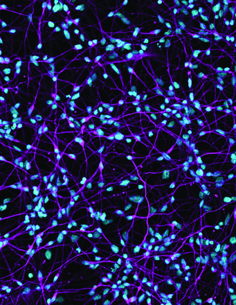
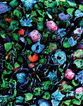
SfN uses scientific imagery in support of its annual meeting, report, and associated products. Listed below is the scientific imagery used each year alongside descriptions and image credits.
Use the following links to navigate to the scientific images you are interested in learning more about.
- 2025 Annual Meeting Science Image Credits
- 2024 Annual Meeting, Products, and Merchandise
- 2023 Annual Meeting
- 2023 Annual Report
- 2022 Annual Meeting
- 2022 Annual Report
Scientific Imagery Archives
2023 Annual Meeting
Description
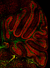
The image shows a postnatal mouse cerebellum stained for Pax2 (green) and NeuN (red), highlighting immature inhibitory interneurons and excitatory granule cells, respectively.
Credited to: Wen Li. Journal of Neuroscience, 29 June 2022, 42 (26) 5130-5143; DOI: https://www.jneurosci.org/content/42/26
View Neuroscience 2023 Scientific Imagery (PDF)
2023 Annual Report
Description
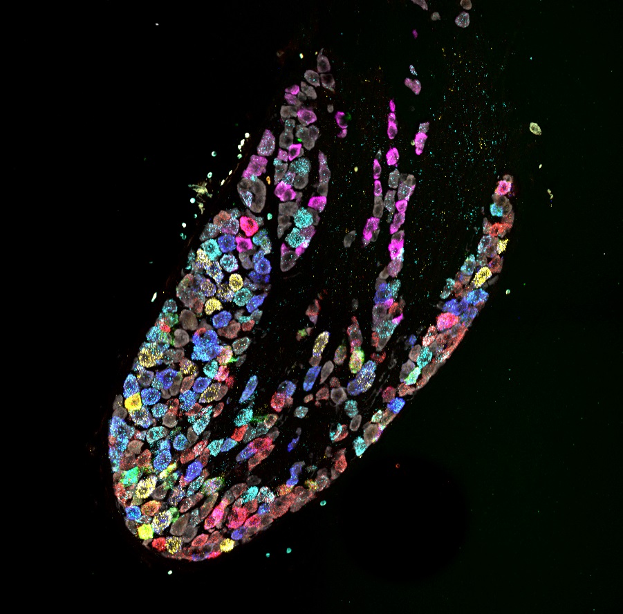
This image shows the emergence of cell type specific gene expression patterns in developing sensory neurons of the vagus nerve located in the nodose ganglion.
Credited to: Meaghan E. McCoy and Anna K. Kamitakahara; eNeuro, 27 March 2023, 10 (4) ENEURO.0511-22.2023; DOI: https://doi.org/10.1523/ENEURO.0511-22.2023
See also: https://blog.eneuro.org/2023/06/snapshots_sensory_cell_types
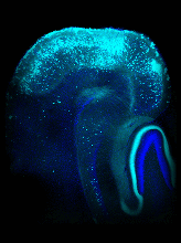
Description
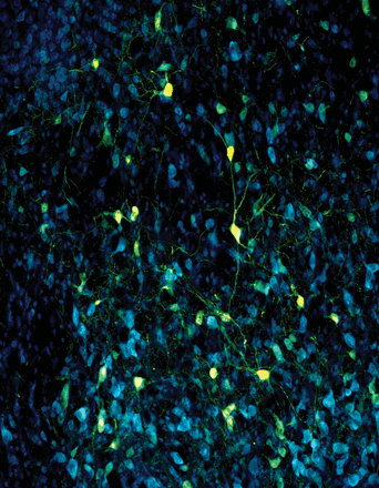
In this confocal image, the projection neurons from the entorhinal cortex are labeled with an antibody against mCherry (cyan). The neurons were targeted using neonatal injection of an adenoassociated virus expressing chemogenetic hM4D(Gi) receptors. This technique allows the chronic silencing of the main excitatory inputs in the hippocampus during postnatal development. For more information, see the article by Feng et al. (pages 4607–4619).
Credited to: Ting Feng, Christian Alicea, Vincent Pham, Amanda Kirk and Simon Pieraut; Journal of Neuroscience, 26 May 2021, 41 (21) 4607-4619; DOI: https://doi.org/10.1523/JNEUROSCI.1207-20.2021
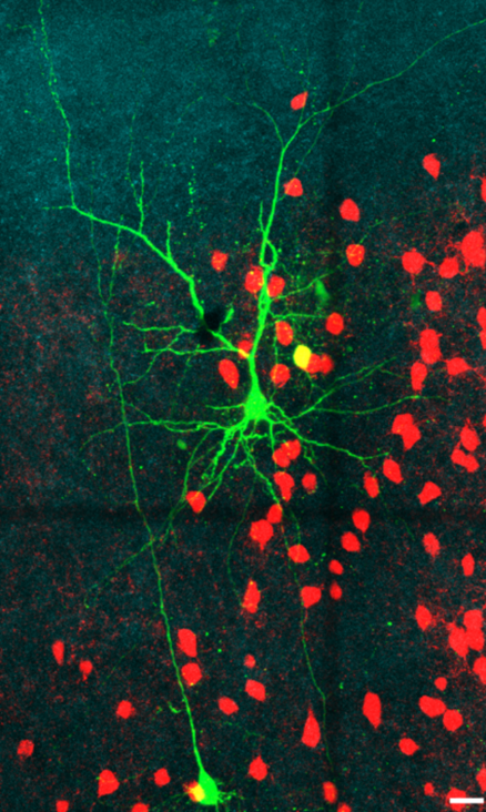
Description
Example of biocytin filled 3 cells (green) and current-clamp traces show characteristic PV+ (red) and Pyr neurons subthreshold responses and suprathreshold AP firing in response to depolarizing current steps (PV+ red, Pyr gray) with ChR2 positive axons (cyan). One PV+ filled with biocytin is shown in yellow, because of overlap of tdtomato labeling.
Credited to: Roman U. Goz and Bryan M. Hooks; eNeuro, 24 April 2023, 10 (5) ENEURO.0488-22.2023; DOI: https://doi.org/10.1523/ENEURO.0488-22.2023
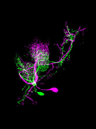
Description
This image shows the lateral antennal lobe tract projection neuron 4 in male (magenta) and female (green) fruit flies. This sexually dimorphic neuron in males helps ensure the proper sequencing of male courtship behaviors. For more information see the article by Tanaka et al. (pages 9732–9741).
Credited to: Nobuaki K. Tanaka, Takashi Hirao, Hikaru Chida and Aki Ejima. Journal of Neuroscience 24 November 2021, 41 (47) 9732-9741; DOI: https://doi.org/10.1523/JNEUROSCI.1168-21.2021
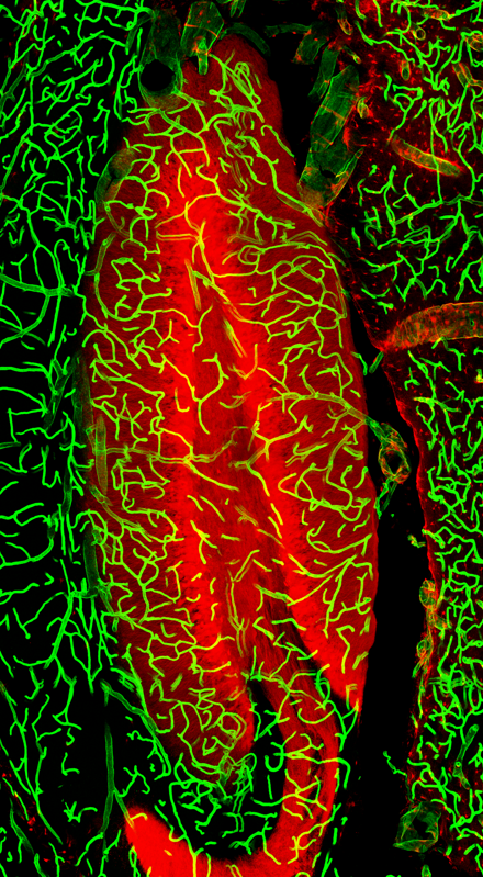
Description
Confocal image of hippocampal dentate gyri showing granule cells expressing tdTomato (red) and blood vessels labeled with DyLight 649-lectin (green).
Credited to: Mary Dusing, Candi L. LaSarge, Angela White, Lilian G. Jerow, Christina Gross, and Steve C. Danzer. eNeuro, 9 February 2023, 10 (2) ENEURO.0340-22.2023; DOI: https://doi.org/10.1523/ENEURO.0340-22.2023
2022 Annual Meeting
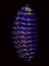
Description
This image shows the expression of miR-263b in a stage-16 Drosophila embryo. Sensory neurons are shown in red, nuclei in blue (DAPI), and expression of the miR-263b driver in green.
Credited to: Marleen Klann, A. Raouf Issa, Sofia Pinho and Claudio R. Alonso. Journal of Neuroscience, 6 October 2021, 41 (40) 8297-8308; DOI: https://doi.org/10.1523/JNEUROSCI.0081-21.2021
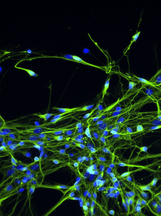
Description
This image depicts cortical neurons derived from human induced pluripotent stem cells, labeled with antibodies against beta-tubulin III (Green). These cells were used to study the epilepsyassocated L1342P genetic variant of Nav1.2 voltage-gated sodium channels. Nuclei were labeled with DAPI (blue).
Credited to: Zhefu Que, Maria I. Olivero-Acosta, Jingliang Zhang, Muriel Eaton, Anke M. Tukker, Xiaoling Chen, Jiaxiang Wu, Junkai Xie, Tiange Xiao, Kyle Wettschurack, Layan Yunis, J. Marshall Shafer, James A. Schaber, Jean-Christophe Rochet, Aaron B. Bowman, Chongli Yuan, Zhuo Huang, Chang-Deng Hu, Darci J. Trader, William C. Skarnes and Yang Yang. Journal of Neuroscience, 8 December 2021, 41 (49) 10194-10208; DOI: https://doi.org/10.1523/JNEUROSCI.0564-21.2021
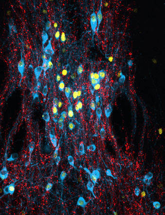
Description
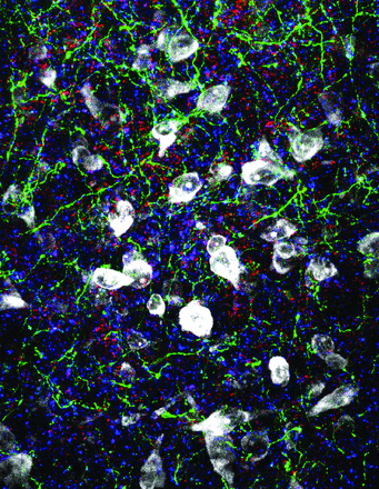
This confocal image shows the innervation of dopaminergic neurons in the caudal linear raphe nucleus by the bed nucleus of the stria terminalis (BNST) and the activation pattern of these neurons during fear memory consolidation. Dopaminergic neurons were co-immunostained with c-Fos (yellow) and tyrosine hydroxylase (cyan), while hM3Dq-expressing BNST axons were visualized by anti-RPF immunostaining (red). Activation of GABAergic neurons of BNST during fear conditioning or memory consolidation resulted in an enhanced cue-related fear recall.
Credited to: Biborka Bruzsik, Laszlo Biro, Dora Zelena, Eszter Sipos, Huba Szebik, Klara Rebeka Sarosdi, Orsolya Horvath, Imre Farkas, Veronika Csillag, Cintia Klaudia Finszter, Eva Mikics and Mate Toth. Journal of Neuroscience 3 March 2021, 41 (9) 1982-1995; DOI: https://doi.org/10.1523/JNEUROSCI.1944-20.2020
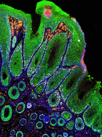
Description
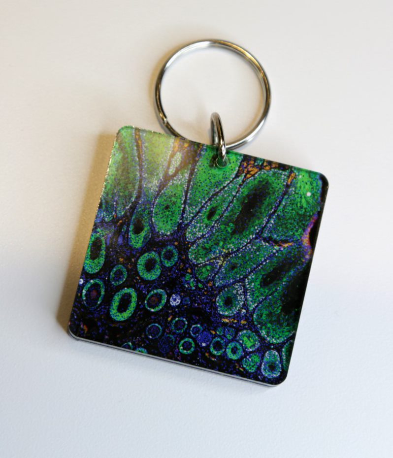
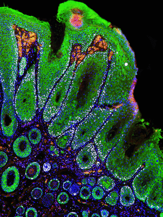
This image shows keratinocytes (green), proliferating cells (white), inflammatory cells (red), and cell nuclei (blue) associated with a skin tumor in a one-year-old mouse lacking PTEN, a protein that regulates cell growth and proliferation. Although loss of PTEN is associated with several cancers, it can also promote axon regeneration in the CNS and PNS.
Credited to: Sofia Meyer zu Reckendorf, Diana Moser, Anna Blechschmidt, Venkata Neeha Joga, Daniela Sinske, Jutta Hegler, Stefanie Deininger, Alberto Catanese, Sabine Vettorazzi, Gregor Antoniadis, Tobias Boeckers and Bernd Knöll. Journal of Neuroscience 23 March 2022, 42 (12) 2474-2491; DOI: https://doi.org/10.1523/JNEUROSCI.1305-21.2022
Description
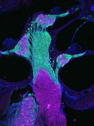
This image shows a cross-section of the cochlea within the inner ear of a reporter mouse. P2×7 receptor-expressing satellite glial cells and Schwann cells are labelled with GFP (green) and spiral ganglion neurons are immunolabeled for bIII-tubulin (magenta).
Credited to: Silvia Prades, Gregory Heard, Jonathan E. Gale, Tobias Engel, Robin Kopp, Annette Nicke, Katie E. Smith and Daniel J. Jagger. Journal of Neuroscience 24 March 2021, 41 (12) 2615-2629; DOI: https://doi.org/10.1523/JNEUROSCI.2240-20.2021
Description
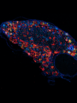
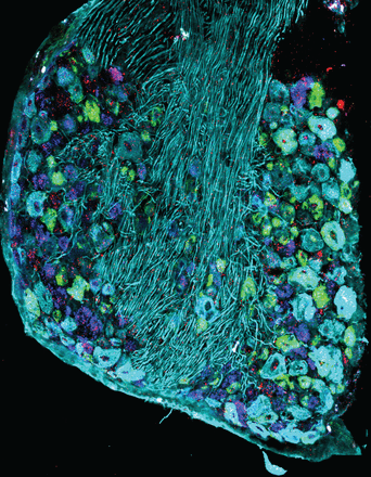
This image shows a section from a mouse dorsal root ganglion stained for Interferon Alpha and Beta Receptor Subunit 1 (mRNA; red) along with the sensory neuron markers calcitonin gene-related peptide (mRNA; green), purinergic receptor P2X3 (mRNA; blue) and Neurofilament-200 (protein; cyan). Interferon receptors were robustly expressed in sensory neurons in the dorsal root ganglion and their activation produces pain via an eIF4E-dependent translation mechanism.
Credited to: Paulino Barragán-Iglesias, Úrzula Franco-Enzástiga, Vivekanand Jeevakumar, Stephanie Shiers, Andi Wangzhou, Vinicio Granados-Soto, Zachary T. Campbell, Gregory Dussor and Theodore J. Price. Journal of Neuroscience 29 April 2020, 40 (18) 3517-3532; DOI: https://doi.org/10.1523/JNEUROSCI.3055-19.2020
Description
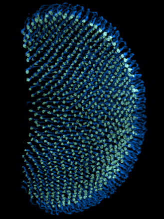
This image shows the axonal projections of R7 photoreceptors in the Drosophila medulla (axons in blue, axonal terminals in green). The stereotypy of these axonal projections makes them a useful model system for acquiring quantitative data on sporadic and progressive axonal degeneration of photoreceptor cells.
Credited to: Mélisande Richard, Karolína Doubková, Yohei Nitta, Hiroki Kawai, Atsushi Sugie and Gaia Tavosanis. Journal of Neuroscience 15 June 2022, 42 (24) 4937-4952; DOI: https://doi.org/10.1523/JNEUROSCI.2115-21.2022
Description
Three-dimensional deconvolved fluorescence image of cultured hippocampal neurons in which cellular components involved in synaptic activity, activity-dependent transcription, and synaptic plasticity are labeled. The presynaptic protein synapsin I (green) and the dendritic microtubule-associated protein MAP2 (blue) are labeled with antibodies, ribosomes (red) are labeled with NeuroTrace Nissl stain, and nuclear DNA (yellow) is labeled with Hoechst dye. Synaptic activity regulates gene transcription via activity-sensitive transcription factors, including CaRF (calcium-response factor).
Credited to: Ronan Chereau, Kelli A. McDowell, Ashley N. Hutchinson, Sarah J.E. Wong-Goodrich, Matthew M. Presby, Dan Su, Ramona M. Rodriguiz, Krystal C. Law, Christina L. Williams, William C. Wetsel and Anne E. West. Journal of Neuroscience 2 June 2010, 30 (22) 7453-7465; DOI: https://doi.org/10.1523/JNEUROSCI.3997-09.2010
Description
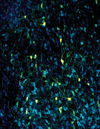
This image shows ventral striatal neurons from a rodent brain. The cell bodies are stained in blue using a fluorescent NISSL stain (NeuroTrace). A light sensitive virus for optogenetics was transduced into neurons shown in yellow. Neurons in this area are involved in scaling fear to the degree of threat and in rapid discrimination of uncertain threat and safety.
Credited to: Madelyn H. Ray, Alyssa N. Russ, Rachel A. Walker and Michael A. McDannald. Journal of Neuroscience 10 June 2020, 40 (24) 4750-4760; DOI: https://doi.org/10.1523/JNEUROSCI.0299-20.2020
Description
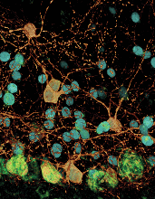
This confocal image depicts the morphological details of cerebellar molecular layer interneurons (MLIs). Single MLIs are lighted up in orange using Cre-dependent fluorescent markers. MLI nuclei are labelled in cyan using nuclei-targeted GFP. Cerebellar Purkinje neuron somas are labelled by NeuroTrace and shown in green.
Credited to: Candace H. Carriere, Wendy Xueyi Wang, Anson D. Sing, Adam Fekete, Brian E. Jones, Yohan Yee, Jacob Ellegood, Harinad Maganti, Lola Awofala, Julie Marocha, Amar Aziz, Lu-Yang Wang, Jason P. Lerch and Julie L. Lefebvre. Journal of Neuroscience 4 November 2020, 40 (45) 8652-8668; DOI: https://doi.org/10.1523/JNEUROSCI.1636-20.2020
Description
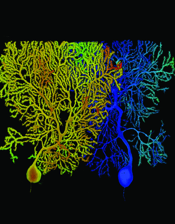
This image shows the dendritic trees of two Purkinje cells from wild-type (left, orange) and mutant mice lacking the alpha- and gamma-Protocadherin gene clusters (Pcdhs) (right, blue). Purkinje cells were labelled with fluorophores, and shown as confocal projections with surface rendering and color to illustrate depth. Pcdhs promote dendrite self-avoidance to ensure that Purkinje dendrites are evenly arranged across the arbor, with minimal overlaps between branches.
Credited to: Julie Marocha. Journal of Neuroscience 14 March 2018, 38 (11) 2713-2729; DOI: https://www.jneurosci.org/content/38/11/2713?iss=11
2022 Annual Report
Description
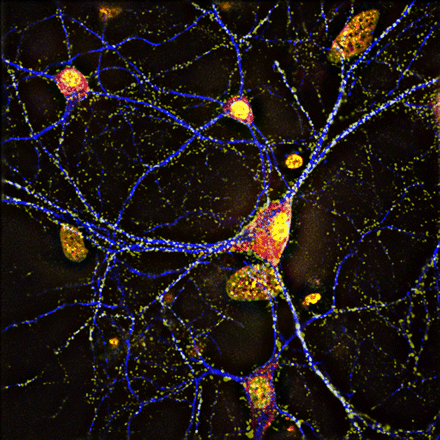
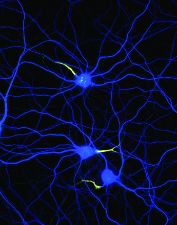
This image shows cultured hippocampal neurons labeled for dendrites (MAP2, blue), and for the axon initial segment (yellow is an overlay of neurofascin-186, green and β4-spectrin, red).
Credited to: Christophe Leterrier. Journal of Neuroscience 28 February 2018, 38 (9) 2135-2145; DOI: https://doi.org/10.1523/JNEUROSCI.1922-17.2018
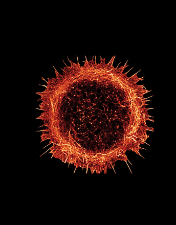
Description
This STORM image shows a rat hippocampal neuron stained for actin 24 h after plating in culture. It is still in the first stage of growth, with a peripheral actin-rich lamellipodium but no neurites. Some of the actin spikes will become neurites.
Credited to: Christophe Leterrier. Journal of Neuroscience 6 January 2021, 41 (1) 11-27; DOI: https://doi.org/10.1523/JNEUROSCI.2872-20.2020
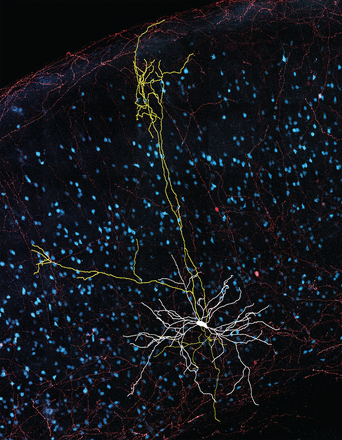
Description
This image shows the axon (yellow), soma and dendrites (white) of a somatostatin-expressing (cyan) neuron in mouse auditory cortex. A retrograde tracer (magenta) revealed that this neuron sends a long-range projection to the ipsilateral lateral amygdala.
Credited to: Alice Bertero, Paul Luc Caroline Feyen, Hector Zurita and Alfonso junior Apicella. Journal of Neuroscience 23 October 2019, 39 (43) 8424-8438; DOI: https://doi.org/10.1523/JNEUROSCI.1515-19.2019
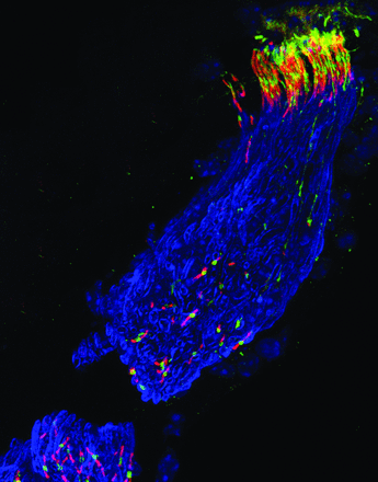
Description
This image shows mature cochlear heminodes beneath hair cells and nodes of Ranvier within osseous spiral lamina in adult mouse auditory nerve. The nodes and their flanking paranodes were immunolabeled for neuronal cell adhesion molecule (NrCAM, green) and contactin 1 (Cntn1, red), respectively. Myelin of the auditory nerve (following the heminodes) was detected by immunolabeling for myelin basic protein (MBP, blue; nuclei were counterstained with DAPI also in blue). The integrity of myelin and nodal structures in the cochlea is needed for fast transfer of sound information from the hair cells to the brain.
Credited to: Clarisse H. Panganiban, Jeremy L. Barth, Lama Darbelli, Yazhi Xing, Jianning Zhang, Hui Li, Kenyaria V. Noble, Ting Liu, LaShardai N. Brown, Bradley A. Schulte, Stéphane Richard and Hainan Lang. Journal of Neuroscience 7 March 2018, 38 (10) 2551-2568; DOI: https://doi.org/10.1523/JNEUROSCI.2487-17.2018
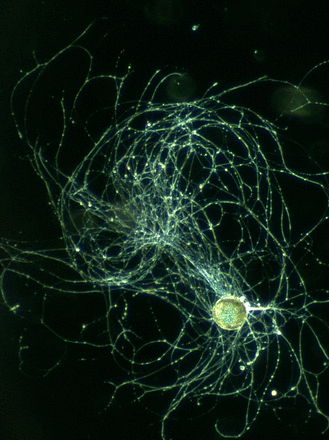
Description
This image shows an isolated B8 motor neuron from an Aplysia californica buccal ganglion. This neuron controls a muscle involved in food grasping and intake during feeding.
Credited to: Yuto Momohara, Curtis L. Neveu, Hsin-Mei Chen, Douglas A. Baxter and John H. Byrne. Journal of Neuroscience 16 February 2022, 42 (7) 1211-1223; DOI: https://doi.org/10.1523/JNEUROSCI.1722-21.2021
Description
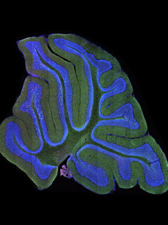
This confocal image of a sagittal section of the mouse cerebellum shows mossy fiber terminals (red, green, yellow) from Clarke's column neurons in the spinal cord, which synapse onto granule cells (blue) in the cerebellum. Sparse labeling of lower thoracic to lumbar Clarke's column neurons (green or yellow) reveals that Clarke's column neurons extensively expand and distribute proprioceptive information across several discrete lobules, allowing parallel processing across several domains.
Credited to: Iliodora V. Pop, Felipe Espinosa, Cheasequah J. Blevins, Portia C. Okafor, Osita W. Ogujiofor, Megan Goyal, Bishakha Mona, Mark A. Landy, Kevin M. Dean, Channabasavaiah B. Gurumurthy and Helen C. Lai. Journal of Neuroscience 26 January 2022, 42 (4) 581-600; DOI: https://doi.org/10.1523/JNEUROSCI.2157-20.2021
Description
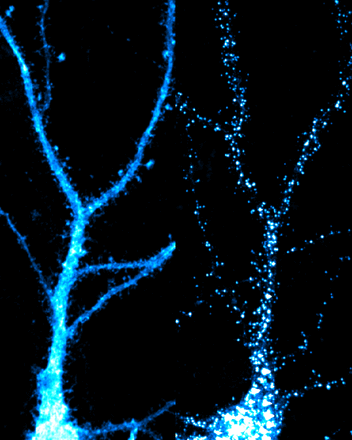
Confocal immunofluorescent image of two hippocampal neurons labeled for HA-tagged Nedd4-1, with and without AMPA treatment. In basal conditions (left), the E3 ubiquitin ligase Nedd4-1 is diffusely localized throughout the soma and dendrites. Upon activation of AMPA receptors (right), Nedd4-1 is rapidly recruited to excitatory synapses within dendritic spines, where it is able to ubiquitinate surface AMPA receptors and manipulate synaptic strength.
Credited to: Samantha L. Scudder, Marisa S. Goo, Anna E. Cartier, Alice Molteni, Lindsay A. Schwarz, Rebecca Wright and Gentry N. Patrick. Journal of Neuroscience 10 December 2014, 34 (50) 16637-16649; DOI: https://doi.org/10.1523/JNEUROSCI.2452-14.2014
Description
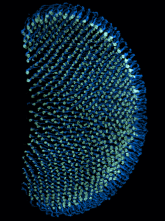
This image shows the axonal projections of R7 photoreceptors in the Drosophila medulla (axons in blue, axonal terminals in green). The stereotypy of these axonal projections makes them a useful model system for acquiring quantitative data on sporadic and progressive axonal degeneration of photoreceptor cells.
Credited to: Mélisande Richard, Karolína Doubková, Yohei Nitta, Hiroki Kawai, Atsushi Sugie and Gaia Tavosanis. Journal of Neuroscience 15 June 2022, 42 (24) 4937-4952; DOI: https://doi.org/10.1523/JNEUROSCI.2115-21.2022
Description
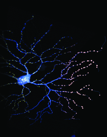
This image shows a retinal ganglion cell that was biolistically labeled in an adult mouse. Cytosolic expression of fluorescent protein tdTomato (blue) reveals cellular morphology, while coexpression of YFP-tagged PSD95 (yellow) labels excitatory postsynaptic sites within the same neuron. The left portion of the image represents the rendered volume of fluorescent signals expressed by the cell, gradually blended with its digitized representation of the dendritic arbor's skeleton (blue lines) and synaptic loci (pink spheres).
Credited to: Yvonne Ou, Rebecca E. Jo, Erik M. Ullian, Rachel O.L. Wong and Luca Della Santina. Journal of Neuroscience 31 August 2016, 36 (35) 9240-9252; DOI: https://doi.org/10.1523/JNEUROSCI.0940-16.2016
Description
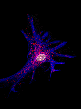
This pseudo-colored photomicrograph depicts mitochondrial (MitoTracker, red) distribution in a primary cultured mouse cortical astrocyte immunostained for GFAP (blue) and nuclei (white).
Credited to: Matthew P. Baier, Raghavendra Y. Nagaraja, Hannah P. Yarbrough, Daniel B. Owen, Anthony M. Masingale, Rojina Ranjit, Megan A. Stiles, Ashley Murphy, Martin-Paul Agbaga, Mohiuddin Ahmad, David M. Sherry, Michael T. Kinter, Holly Van Remmen and Sreemathi Logan. Journal of Neuroscience 3 August 2022, 42 (31) 5992-6006; DOI: https://doi.org/10.1523/JNEUROSCI.2543-21.2022
Description
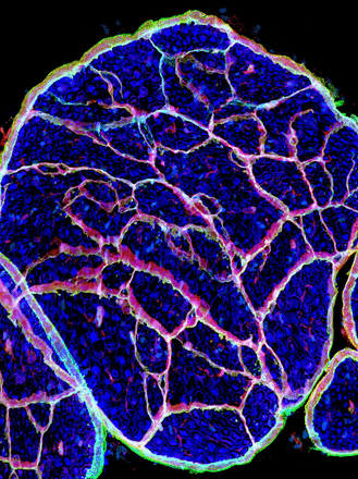
Confocal image of a section of sciatic nerve from an adult mouse lacking Gli-1, a transcription factor expressed in the endoneurial compartment of peripheral nerves independent of its canonical activator, the Hedgehog pathway. Fate mapping with Gli1-Cre (red) labels both the perineurium, which surrounds the nerve exterior, and cells forming minifascicles, structures that aberrantly subdivide these knockout peripheral nerves into multiple small compartments. The nerve is additionally stained for axonal neurofilament (blue) and the glucose transporter Glut-1 (green).
Credited to: Brendan Zotter, Or Dagan, Jacob Brady, Hasna Baloui, Jayshree Samanta and James L. Salzer. Journal of Neuroscience 12 January 2022, 42 (2) 183-201; DOI: https://doi.org/10.1523/JNEUROSCI.3096-20.2021


